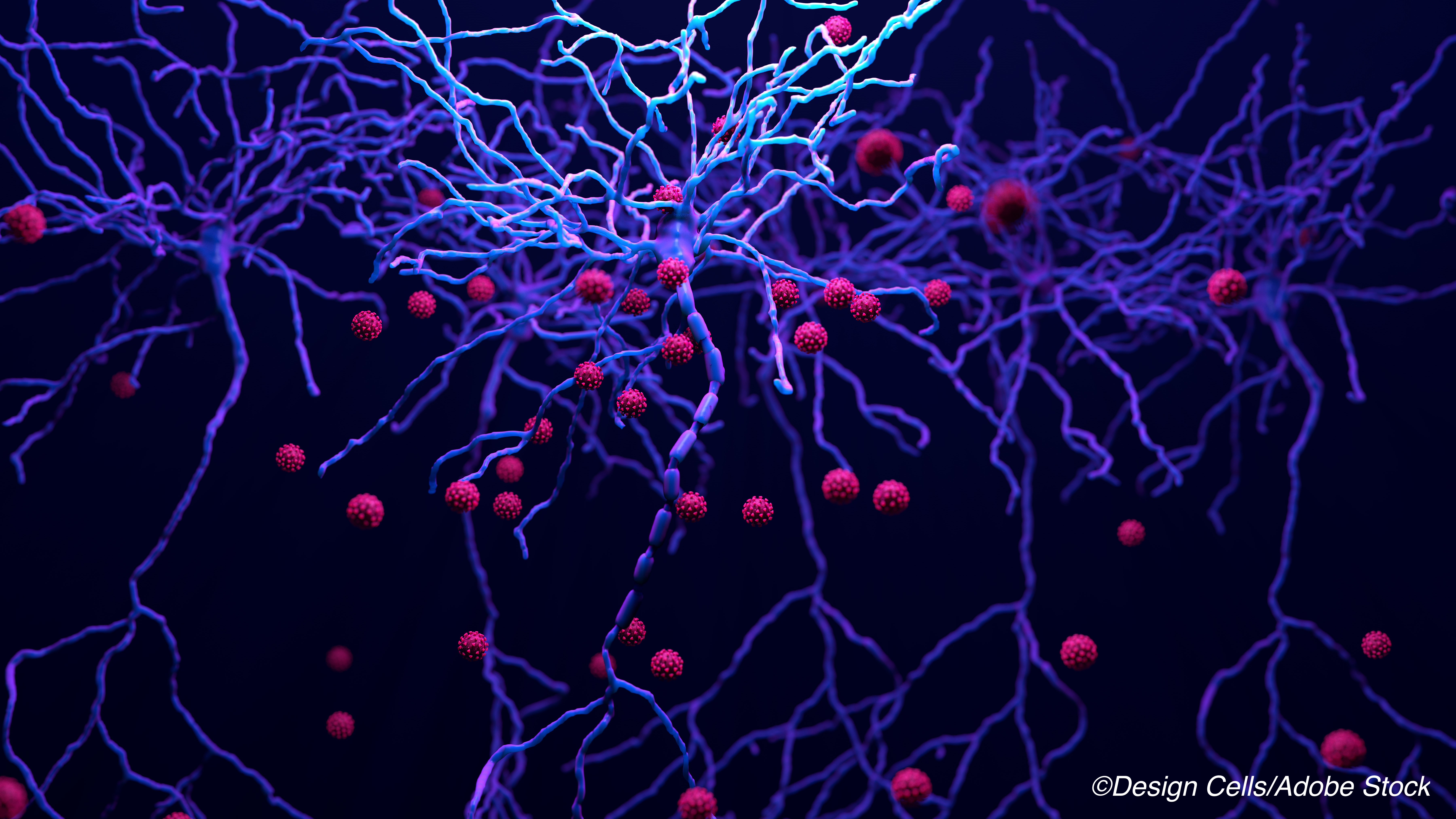
SARS-CoV-2 RNA and proteins were found in brain tissues, but there was no evidence for direct, virally caused neuropathologic change, an autopsy series of 43 Covid-19 patients found.
Quantitative RT-PCR and immunohistochemistry staining showed either SARS-CoV-2 RNA, viral proteins, or both in brain tissue of 53% of patients who died with Covid-19, reported Markus Glatzel, MD, of the University Medical Center Hamburg-Eppendorf in Germany, and coauthors.
“Neuropathological changes in patients with Covid-19 seem to be mild, with pronounced neuroinflammatory changes in the brainstem being the most common finding,” Glatzel and colleagues wrote in Lancet Neurology.
“The neuropathological alterations are most likely to be immune-mediated, and there does not seem to be fulminant virus-induced encephalitis nor direct evidence for SARS-CoV-2-caused central nervous system damage,” they added.
A variable degree of astrogliosis was seen in all patients, with 86% showing astrogliosis in all regions assessed in the autopsies. Diffuse activation of microglia with occasional microglial nodules was pronounced in the brainstem and cerebellum.
While cytotoxic T lymphocytes could be seen in small amounts in the frontal cortex, basal ganglia, and infiltrating meninges, they were most pronounced in the brainstem. The olfactory bulb showed pronounced astrogliosis and microgliosis, but minimal infiltration by cytotoxic T lymphocytes.
“We observed substantial yet highly variable degrees of astrogliosis in all assessed regions,” Glatzel and co-authors noted. “Astrocytes are key regulators of homoeostasis, responding to stimuli through upregulation of GFAP and astroglial hypertrophy. Because astrogliosis occurs in a variety of pre-existing medical conditions, and because critical illness also contributes to astrogliosis, a causal connection to SARS-CoV-2 cannot be drawn at present.”
“Further studies are needed to define how SARS-CoV-2 gains access to the brain, to define the neuroimmune activation, and to describe the distribution of SARS-CoV-2 in the brain,” they added.
In an accompanying editorial, Stephan Frank, PhD, of the University of Basel in Switzerland, noted that “the question of whether the neuropathological alterations in COVID-19 directly result from SARS-CoV-2 brain infection, as opposed to reflecting sequelae of an overstimulated systemic immune response, is of high clinical importance.”
“Whereas the first scenario would support the use of remdesivir or other antivirals, anti-inflammatory modalities appear to be the treatment of choice once damaging immunoinflammatory mechanisms take over,” he wrote. “Teasing apart these fundamentally different scenarios is an ongoing task for neuropathology experts.”
In this study, Glatzel and colleagues conducted autopsies on 43 patients age 51 to 94 who died after PCR-confirmed diagnosis of SARS-CoV-2 infection from March 13 to April 24. Median age was 76 and 37% were women. Pre-existing chronic medical conditions, mainly cardiorespiratory problems, were present in 92% overall, and 30% had pre-existing neurologic illness including neurodegenerative disease or epilepsy.
The mean post-mortem interval was 3.3 days, and 53% showed mild to moderate brain edema, consistent with nonspecific agonal change.
SARS-CoV-2 RNA was detected in frontal lobe tissue in 39% of 23 patients with available samples and in medulla oblongata tissue in 50% of 8 patients with available samples. In eight patients who were untested or tested negative for viral RNA in brain tissue, viral proteins were seen in the medulla oblongata associated with isolated medullary cells and the glossopharyngeal and vagal nerves near their origins from the brainstem.
“We saw a surprisingly uniform presentation of neuropathological findings (i.e., activation of microglia, infiltration with CD8-positive T cells) in our patients, irrespective of the clinical severity of Covid-19 in each case,” Glatzel and co-authors noted.
“Remarkably, SARS CoV-2 presence did not correlate with the severity of neuropathological alterations,” observed Frank. “While the replicative and infective potential of the viral RNS remains unclear, the in-situ detection of SARC-CoV-2 proteins is an important finding as it confirms the presence of the virus in the brain.”
Viral protein expression was confined to the medulla and exiting cranial nerves, Frank pointed out: “Considering the capability of SARS-CoV-2 to infect human gut enterocytes as well as pneumocytes, this finding is of particular interest, warranting future investigations of vagal nerve tissue as a potential viral CNS access route in Covid-19.”
Limitations of the study include a low number of cases, and age and sex-matched controls were not included. Limitations inherent to postmortem studies include varying postmortem intervals and incomplete clinical data about the patient.
In addition, “common Covid-19 treatment modalities, such as invasive ventilation (which might promote cerebral microbleeds) or dexamethasone medication (known to modulate immune responses), have to be considered when interpreting neuropathological findings,” Frank noted.
- SARS-CoV-2 RNA and proteins were found in brain tissues, but there was no evidence for direct, virally caused neuropathologic change, an autopsy series of Covid-19 patients found.
- The replicative and infective potential of viral RNS remains unclear, but in-situ detection of SARS-CoV-2 proteins is an important finding as it confirms the presence of the virus in the brain.
Paul Smyth, MD, Contributing Writer, BreakingMED™
Funding came from German Research Foundation, Federal State of Hamburg, EU (eRARE), German Center for Infection Research (DZIF).
Glatzel has no disclosures.
Frank reports grants from the Botnar Research Centre for Child Health.
Cat ID: 130
Topic ID: 82,130,791,932,580,190,926,130,192,927,151,928,925,934


